Positive and negative toolingArray drawing reviewOptical inspection
Home / Research library

Research library

Read the original tooling studies alongside guides to orientation, cavity images and inspection records.
Array tooling in micrographs

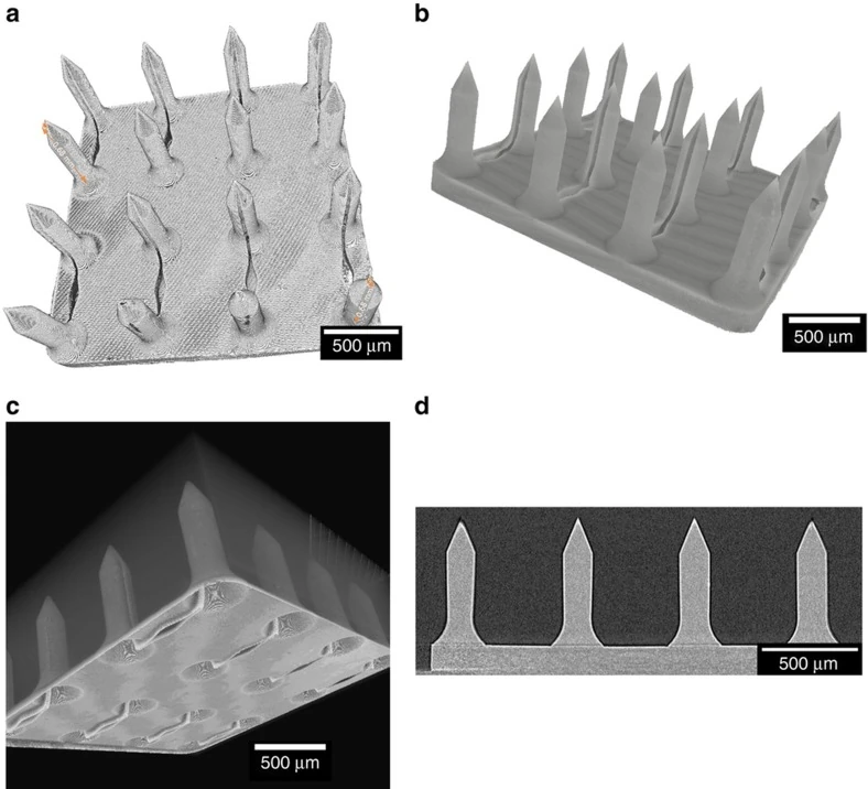
Four micro-CT panels showing a master, a positive replica, an inverse mold and a section through the master.
Master, mold and replica in micro-CTFaraji Rad and colleagues imaged the three physical stages and included a master section for comparison.
Image creditsZahra Faraji Rad, Robert E Nordon, Carl J Anthony, Lynne Bilston, Philip D Prewett, Ji-Youn Arns, Christoph H Arns, Liangchi Zhang and Graham J Davies · Image source · CC BY 4.0 · Original figure proportions and markings retained; converted to WebP.
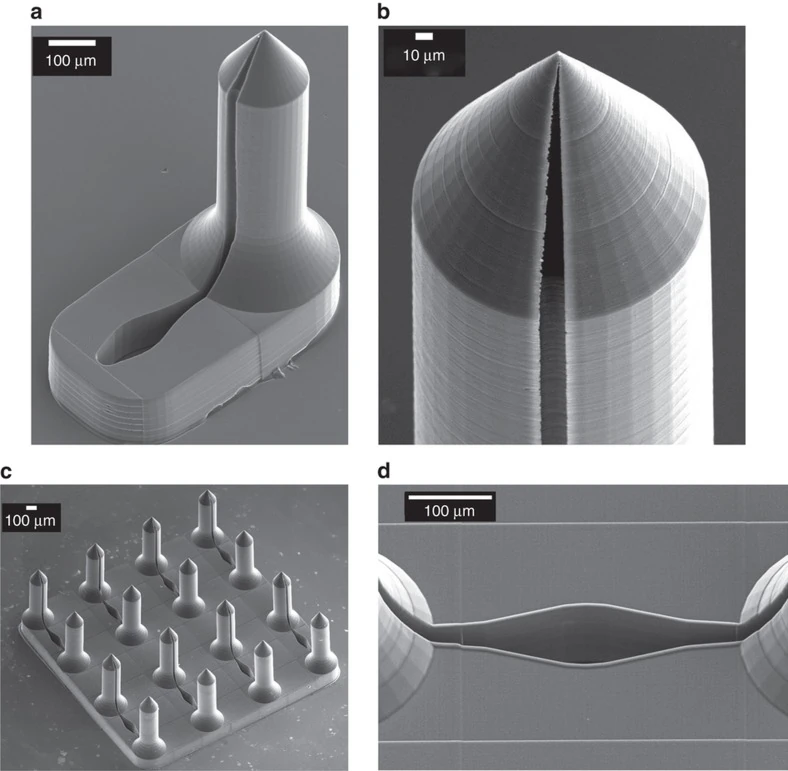
Four grayscale micrographs of a replicated array, an individual feature, its tip and a connecting channel.
Replica array and feature detailThe researchers documented the same replica design at array and feature scale.
Image creditsZahra Faraji Rad, Robert E Nordon, Carl J Anthony, Lynne Bilston, Philip D Prewett, Ji-Youn Arns, Christoph H Arns, Liangchi Zhang and Graham J Davies · Image source · CC BY 4.0 · Original figure proportions and markings retained; converted to WebP.
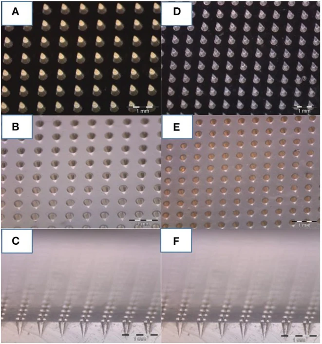
Six optical views showing two master fields, their inverse stamps and side sections through the mold material.
Positive master and negative stampBattisti and colleagues show how a positive field and the corresponding cavity field appear from above and in section.
Image creditsMario Battisti, Raffaele Vecchione, Costantino Casale, Fabrizio A Pennacchio, Vincenzo Lettera, Rezvan Jamaledin, Martina Profeta, Concetta Di Natale, Giorgia Imparato, Francesco Urciuolo and Paolo Antonio Netti · Image source · CC BY 4.0 · Original figure proportions and markings retained; converted to WebP.
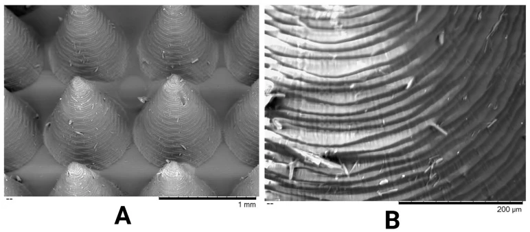
Low-magnification scanning electron image of a conical array beside a higher-magnification image of a single textured flank.
Array spacing and flank textureThe paired magnifications let readers compare spacing across a field with texture on one feature.
Image creditsRutuja N. Meshram and Dimitrios A. Lamprou · Image source · CC BY 4.0 · Original figure proportions and markings retained; converted to WebP.
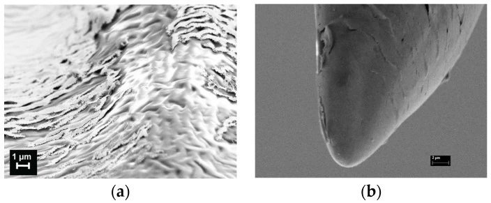
Two scanning electron micrographs show exposed surface texture and the end of a coated microneedle.
Surface texture and apex detailDardano and colleagues examined the surface and apex in separate microscope views.
Image creditsPrincipia Dardano, Alessandro Caliò, Vincenza Di Palma, Maria Fortuna Bevilacqua, Andrea Di Matteo and Luca De Stefano · Image source · CC BY 4.0 · Original figure proportions and markings retained; converted to WebP.
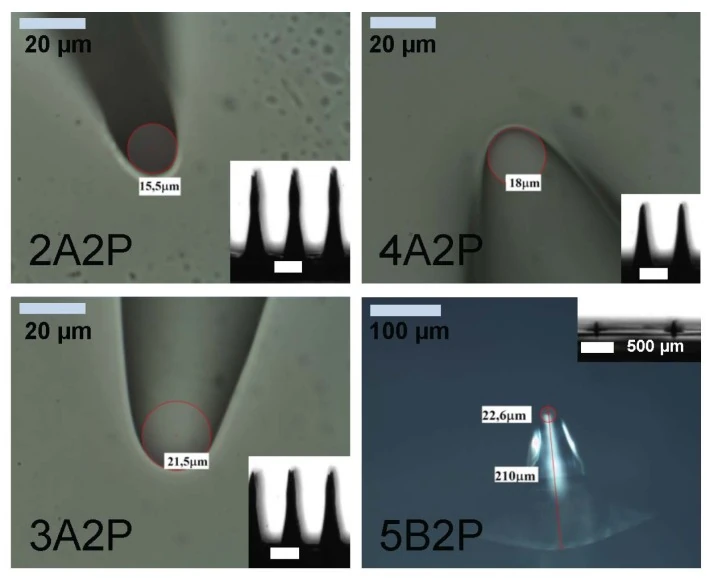
Optical images of four specimens with marked tip contours, profile insets and scale bars.
Annotated tip profilesThese annotations show exactly where the authors read a tip dimension on the pictured specimens.
Image creditsPrincipia Dardano, Alessandro Caliò, Vincenza Di Palma, Maria Fortuna Bevilacqua, Andrea Di Matteo and Luca De Stefano · Image source · CC BY 4.0 · Original figure proportions and markings retained; converted to WebP.
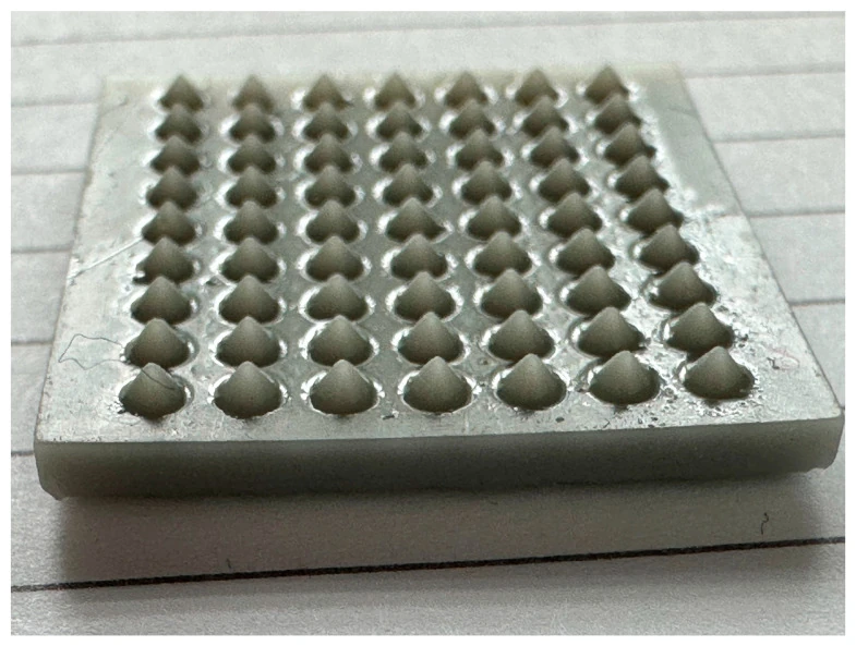
Angled specimen photograph showing a rectangular support and the full field of raised features.
Array on its rectangular supportAldawood and colleagues show the field and its supporting edge in the same view.
Image creditsFaisal Khaled Aldawood, Abhay Andar and Salil Desai · Image source · CC BY 4.0 · Original figure proportions and markings retained; converted to WebP.
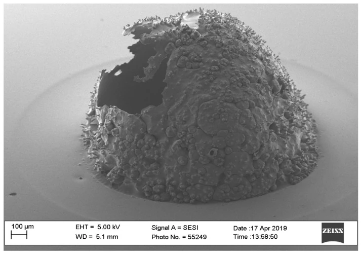
Scanning electron micrograph of a single irregular raised feature with an opening and a textured surface.
Irregular feature under SEMThe visible contour and opening give a reviewer specific features to mark when requesting a second view.
Image creditsFaisal Khaled Aldawood, Abhay Andar and Salil Desai · Image source · CC BY 4.0 · Original figure proportions and markings retained; converted to WebP.
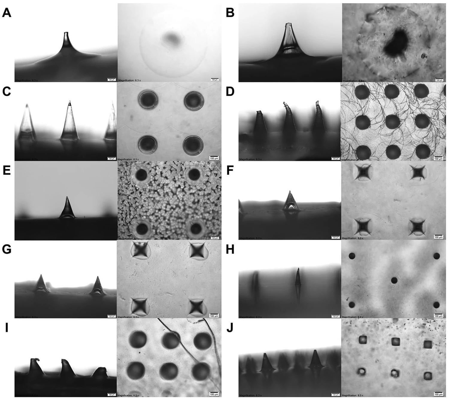
Paired lateral and top microscope views of ten letter-coded patch specimens with scale bars.
Side and top views of coded specimensLee and colleagues kept each specimen code alongside its two microscope views.
Image creditsJing Yi Lee, Shi Hui Dong, Keng Wooi Ng and Choon Fu Goh · Image source · CC BY 4.0 · Original figure proportions and markings retained; converted to WebP.

Guide library

Sources