Optical inspection of microneedle arrays
Request microscope views that connect a local detail to the correct sample, field position and approved drawing.

Request microscope views that connect a local detail to the correct sample, field position and approved drawing.
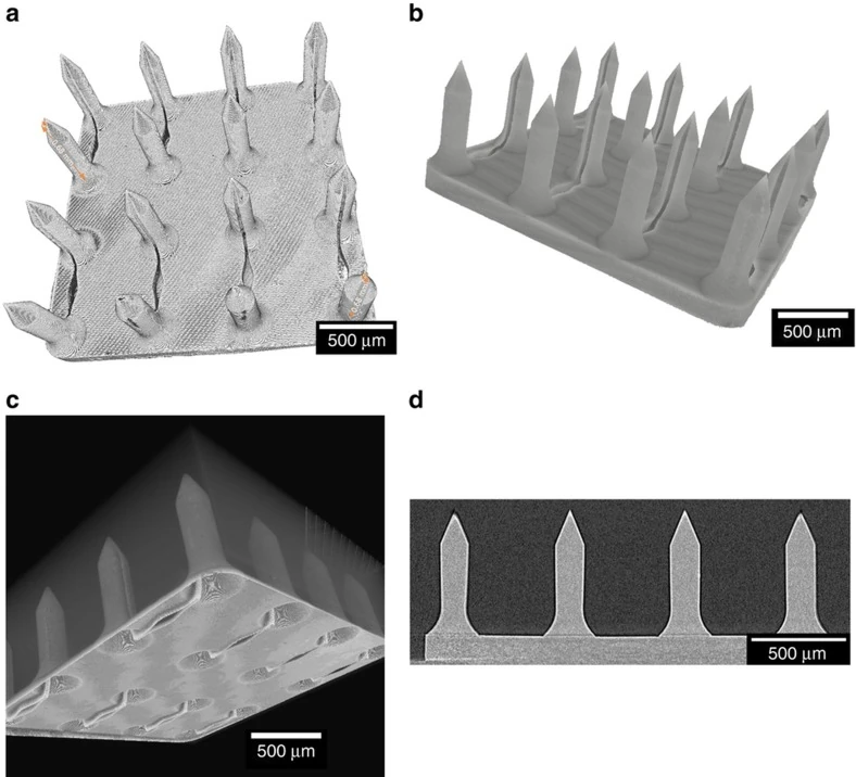
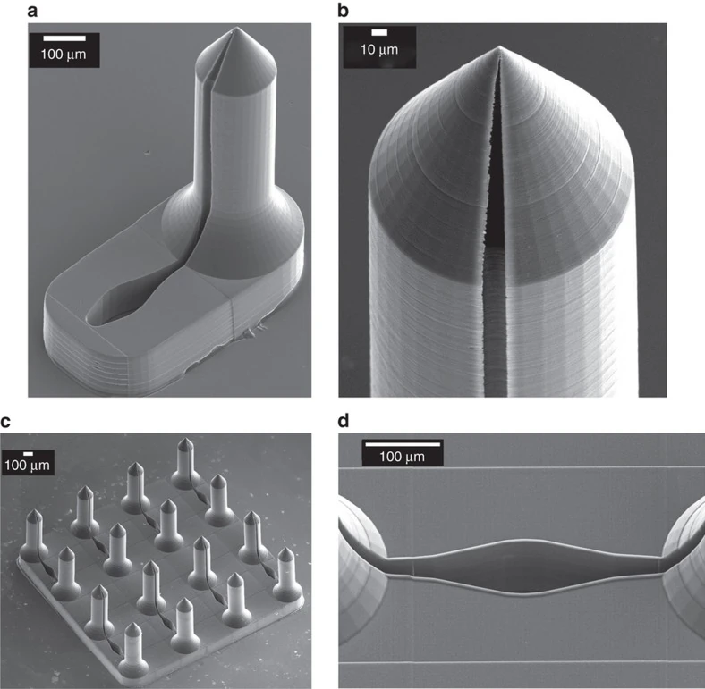
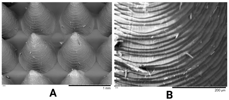
Request microscope views that connect a local detail to the correct sample, field position and approved drawing.
Compare the master, negative cavity and replicated array to investigate a visible difference during a cosmetic tooling review.
Keep the approved master, sample revision and repeat-order scope connected when a buyer orders cosmetic patch tooling again.
Prepare an RFQ for an array drawing review, a positive master, a negative template or a matched cosmetic patch sample.
Match summary values to measured positions and compare edges, corners and center fields across supplier sample reports.
Separate surface texture observations from dimensional form when reviewing master tooling inspection reports and photographs.
Build a first-article file linking a delivered tool, a representative sample and itemized inspection evidence to the approved drawing.
No matching guides. Try a different word.